
410 MHz vs 7 MHz Transducer Frequency: Dermal Energy Attenuation and Ultherapy Depth Physics
The global microfocused ultrasound (MFU-V) and non-invasive skin tightening market is expanding rapidly, driven by multi-layered treatment protocols targeting the 3.0 mm dermis and 4.5 mm SMAS layers. While recent biomedical research introduces ultra-high…
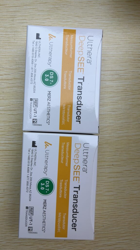
The global microfocused ultrasound (MFU-V) and non-invasive skin tightening market is expanding rapidly, driven by multi-layered treatment protocols targeting the 3.0 mm dermis and 4.5 mm SMAS layers. While recent biomedical research introduces ultra-high frequencies like 410 MHz for cellular-scale imaging, clinical lifting remains anchored in the 4–10 MHz range. For medical aesthetic clinics, dermatology practices, and B2B procurement managers, understanding the core physics of transducer frequency, beam geometry, and dermal energy attenuation is essential to optimizing clinical outcomes, managing consumable budgets, and evaluating new versus certified pre-owned (CPO) device inventory.
As a specialized B2B medical aesthetic integration platform, ALLWILL supports global clinics by supplying authentic Ulthera DS series cartridges, providing standardized testing through its Smart Center network, and offering tailored utilization-based payback models to ensure predictable ROI and safe, reproducible energy delivery.
What is Transducer Frequency in MFU-V?
Transducer frequency in MFU-V describes the number of ultrasound pressure cycles per second, measured in megahertz (MHz), and directly controls wavelength, spatial resolution, focal beam geometry, and the rate of dermal energy attenuation in tissues during non-invasive treatments.
The Core Technical Dilemma: Frequency, Depth, and Clinical Uncertainty
Traditional high-intensity focused ultrasound (HIFU) platforms often lack real-time visualization and rely on single-frequency configurations or unverified depth estimates. This creates a high degree of clinical uncertainty. When therapeutic frequencies are uncalibrated or applied blindly without real-time imaging guidance, energy deposition layers frequently deviate from actual anatomical structures.
If a transducer frequency is too low or miscalibrated for deep targeting, the energy focus can overshoot the superficial muscular aponeurotic system (SMAS) and strike periosteal bone interfaces, causing severe pain and potential nerve irritation. Conversely, if the frequency is too high for the intended deep target, energy is prematurely absorbed and scattered in the superficial dermis, causing near-field heating and superficial burns while leaving the deep foundational layers completely uncorrected.
This anatomical mismatch leads to distinct clinical failures:
-
If a 3.0 mm dermal remodeling pass shifts superficially to 2.0 mm due to poor coupling or unverified beam geometry, patients experience temporary erythema and epidermal swelling without real-time collagen synthesis or long-term lifting.
-
If a 4.5 mm SMAS pass fails to focus precisely on the fascial plane and instead scatters within complex subcutaneous fat-muscular boundaries, treatment reproducibility drops sharply, compromising the clinic’s clinical safety margins.
-
In areas with thin tissue layers or prominent bony contours, such as the peri-orbital and forehead regions, the absence of real-time acoustic feedback forces practitioners to guess tissue thickness, resulting in uneven energy distribution, accelerated transducer wear, and unstable outcomes.
While experimental frequencies like 410 MHz offer unprecedented cell-scale resolution in laboratory settings, their extreme attenuation rates make them completely unusable for structural tightening. Misunderstanding the physics of the “higher frequency equals better device” myth can lead clinics to adopt unverified platforms, resulting in rising consumable costs, unpredictable side effects, and diminished patient satisfaction.
Dermal Acoustic Impedance and Energy Attenuation Physics
Acoustic impedance governs how ultrasound waves behave when crossing boundaries between different soft tissue layers. Significant differences in acoustic impedance at the epidermis-dermis, dermis-fat, fat-SMAS, and tissue-bone interfaces trigger partial reflections and energy scattering. Higher therapeutic frequencies are particularly susceptible to this energy loss.
In soft human tissue, acoustic attenuation increases approximately linearly with frequency. Higher frequency waves experience rapid absorption and scattering per millimeter of tissue traveled, requiring a precise engineering trade-off between target depth and transducer selection.
-
10 MHz Transducers (DS 10-1.5 / DS 10-1.5N): These cartridges exhibit high attenuation per millimeter. They are engineered exclusively for shallow targets where the tissue path length is short, providing sharp, localized thermal coagulation points (TCPs) at a 1.5 mm depth without over-penetrating into underlying fat.
-
7 MHz Transducers (DS 7-3.0 / DS 7-4.5): These cartridges experience lower attenuation per millimeter compared to 10 MHz, allowing sufficient acoustic energy to penetrate deeper into the reticular dermis (3.0 mm) and the SMAS layer (4.5 mm) to anchor structural lifting protocols.
To put this relationship into perspective: ultrasound intensity attenuates as much traveling through just 0.5 cm of soft tissue at 7 MHz as it does traveling through a full 1.0 cm at 3.5 MHz. Every doubling of frequency incurs a steep energy penalty at deeper layers.
Technical Specifications and Beam Geometry Comparison
| Technical Indicator | 410 MHz High-Frequency Transducer | 7 MHz Ulthera Transducer (DS 7-3.0 / DS 7-4.5) | 10 MHz Ulthera Transducer (DS 10-1.5 / DS 10-1.5N) |
| Primary Clinical Application | Ultra-high resolution experimental acoustic microscopy and cellular research | Non-invasive face, neck, and submental structural lifting via MFU-V | Superficial dermal remodeling, fine line correction, and textural refinement |
| Target Focal Depth | Within a few hundred micrometers of the tissue surface | 3.0 mm (deep dermis) and 4.5 mm (SMAS layer) | 1.5 mm (superficial papillary and upper reticular dermis) |
| Wavelength & Spatial Resolution | Cellular-level resolution; wavelength approx. 3.7 μm | Structural tissue-level resolution; wavelength in hundreds of microns | Fine tissue structural resolution for superficial dermal boundaries |
| Beam Width at Focus | Extremely narrow, micro-scale focal point | Approximately 0.27 mm to 0.29 mm | Approximately 0.24 mm to 0.25 mm |
| System Maximum Power Output | Restricted to milliwatt levels to avoid immediate cell lysis | Rated up to 65 W at the system level (Energy under 3 J per shot) | Rated up to 15 W at the system level (Energy under 1.5 J per shot) |
| Tissue Attenuation Rate | Extemely rapid; total energy dissipation in near-surface layers | Balanced attenuation; optimized for deep structural penetration | Accelerated attenuation; restricted to superficial paths to protect deep tissue |
| Visualization & Imaging Integration | Research-grade microscopic visualization | Integrated DeepSEE B-mode real-time imaging up to 8 mm depth | Integrated DeepSEE B-mode real-time imaging for superficial layering |
Multifaceted Dimensions of MFU-V Frequency and Depth
Frequency and Wavelength: Resolution vs. Focal Size
Higher frequencies produce shorter acoustic wavelengths, resulting in smaller focal volumes and superior spatial resolution. A 410 MHz transducer operates with a wavelength of roughly 3.7 μm, creating micro-zones suited for single-cell evaluation but incapable of generating the macrothermal tissue contraction required for clinical lifting. Conversely, 7–10 MHz MFU-V transducers maintain wavelengths scaled to produce precise, stable thermal coagulation points across several millimeters, striking the ideal therapeutic balance for collagen synthesis.
Frequency and Penetration: The Classic Trade-Off
Acoustic energy absorption is directly proportional to frequency. This relationship dictates why lower therapeutic frequencies are mandatory for deep structural targets. In regional nerve blocks and musculoskeletal imaging, transducers operating above 7 MHz are restricted to viewing superficial structures within 2–3 cm, while 3–5 MHz variations are reserved for deeper 4–5 cm volumes. MFU-V clinical protocols mirror this exact physical law, limiting deep 3.0 mm and 4.5 mm fascial targeting to 7 MHz and 4 MHz arrays to prevent complete near-field energy exhaustion.
Frequency and the Thermal Safety Window
The goal of MFU-V is to elevate localized target tissue to a precise thermal window of 60–70°C. This temperature triggers immediate triple-helix collagen denaturation and long-term neocollagenesis while leaving the surrounding epidermis unscathed. A regulated 7–10 MHz frequency profile, combined with real-time DeepSEE visualization, ensures that this thermal safety window is accurately positioned. Utilizing an extreme frequency like 410 MHz would concentrate thermal energy entirely within the upper micrometers of the epidermis, leading to surface charring, severe pain, and zero structural lift.
Clinical Protocol Design and Cross-Channel Multi-Depth Layering
As MFU-V indications expand from mid-face lifting to large-area body contouring—including the décolleté, abdomen, and upper arms—clinics must master multi-depth cartridge sequencing. Rather than relying on a single frequency, advanced protocols stack multiple transducers to treat the tissue volume hierarchically.
-
Deep Structural Layering (4.5 mm Depth): Treatment begins with the DS 7-4.5 cartridge. Operating at 7 MHz, it penetrates the deep subcutaneous boundaries to deliver targeted thermal coagulation points directly into the SMAS layer, establishing a structural foundation.
-
Intermediate Dermal Layering (3.0 mm Depth): The operator transitions to the DS 7-3.0 cartridge. Maintaining a 7 MHz frequency focused at 3.0 mm, this pass targets the deep reticular dermis and the fat-dermis interface to stimulate neocollagenesis, tighten the intermediate structural matrix, and improve overall skin compliance.
-
Superficial Refinement Layering (1.5 mm Depth): The protocol concludes with a 10 MHz cartridge, such as the DS 10-1.5 or its narrow-footprint variant, the DS 10-1.5N. This superficial pass addresses fine lines, epidermal laxity, and localized textural irregularities in highly sensitive zones like the peri-orbital and décolleté regions.
Step-by-Step Guide: Executing a Precise Frequency-Depth Synergistic Protocol
-
Analyze Anatomical Profiles and Target Layers: Assess the patient’s age, skin thickness, and subcutaneous fat distribution via digital palpation and diagnostic imaging to determine whether the clinical priority is deep SMAS lifting, dermal tightening, or superficial texture refinement.
-
Configure the Transducer Matrix: Select the optimal cartridge combination based on the target zone. For comprehensive full-face and neck protocols, prepare a stacked sequence containing the DS 7-4.5 for fascial anchoring, the DS 7-3.0 for deep dermal matrix remodeling, and the DS 10-1.5N for delicate superficial zones.
-
Verify Tissue Layering via Real-Time Visualization: Apply the ultrasound transducer and utilize the integrated DeepSEE B-mode imaging system to visualize anatomical structures up to 8 mm below the skin surface. Verify acoustic coupling, measure epidermal-dermal thickness, and confirm the exact location of bone boundaries to ensure the therapeutic focal zone bypasses major nerves or vascular structures.
-
Program Energy Delivery and Line Densities: Adjust system power settings to deliver the precise thermal dose required to achieve a localized 60–70°C target effect. Cross-reference treatment line densities with the clinic’s consumable budget and the specific cartridge specifications to optimize clinical efficacy and shot efficiency.
-
Execute Orthogonal and Overlapping Vector Lines: Deliver energy lines in a structured, cross-hatched grid pattern. Ensure that the 4.5 mm and 3.0 mm thermal coagulation points form a interconnected, multi-dimensional grid that lifts and tightens the tissue layers uniformly without over-treating localized areas.
-
Monitor Post-Treatment Response and Mid-Lifecycle Adaptation: Schedule comprehensive follow-up evaluations at 60 and 90 days to track collagen remodeling, volumetric lifting, and skin elasticity. Utilize these clinical outcomes to refine future multi-depth transducer selections and customize maintenance touch-up sessions.
Revenue Generation, Asset Optimization, and Procurement Risk Mitigation
In commercial medical aesthetics, the operational challenge centers on selecting a balanced transducer inventory that maximizes patient throughput while controlling consumable overhead. Because every authentic MFU-V cartridge has a finite lifespan governed by its hardcoded shot limits, a clinic’s financial performance depends on precise line-density planning, routine maintenance, and strategic asset procurement.
Clinics must balance capital expenditure by evaluating new versus certified pre-owned (CPO) consoles. While CPO systems offer lower initial acquisition costs, operators must carefully audit remaining cartridge shot counts, software compatibility, and historical service logs to accurately calculate the real cost per treatment line.
To safeguard investments and ensure compliance, biomedical engineers (BMEs) and clinic managers should implement a standardized technical maintenance checklist:
| Item | Maintenance Focus | Why It Matters for 10 MHz vs. 7 MHz | Recommended Action and Interval |
| 1 | Acoustic Membrane Integrity | 10 MHz cartridges operate closer to the epidermis; any membrane delamination or wrinkling increases the risk of superficial burns, acoustic refraction, and coupling failure. | Visually inspect the cartridge membrane prior to every treatment session; immediately discard units showing discoloration or punctures. |
| 2 | Handpiece and Cable Assemblies | Cable strain, micro-fractures, or connector play can disrupt uniform pulse delivery across all high-frequency channels. | Perform weekly physical inspections of cables for kinks or shielding tears; verify connector seating. |
| 3 | Thermal Monitoring Controls | Prolonged usage of 10 MHz cartridges can accelerate near-field thermal accumulation due to higher superficial tissue absorption coefficients. | Verify the functionality of automatic thermal safety lockouts during monthly system diagnostics; pause treatment if the probe housing feels warm. |
| 4 | Shot-Count Logging and Allocation | Deep 7 MHz cartridges bear the heaviest workload in structural lifting protocols and can silently approach end-of-life limits mid-treatment. | Implement automated shot tracking within the clinic’s electronic medical record (EMR) system; order replacements when capacity reaches 15% remaining life. |
| 5 | B-Mode Image Calibration (DeepSEE) | High-contrast visualization is mandatory to confirm the correct target depth before discharging therapeutic 7 MHz or 10 MHz energy pulses. | Conduct monthly calibration checks utilizing specialized tissue phantoms; immediately address image noise, artifacts, or banding. |
| 6 | Firmware and Software Alignment | System software updates frequently optimize pulse duration parameters, acoustic safety margins, and transducer recognition tables. | Annually audit console software versions; ensure CPO hardware is flashed to match current regional regulatory standards. |
| 7 | Sanitization and Disinfection Protocols | Corrosive chemical solvents can cloud optical acoustic windows, degrade watertight seals, and permanently alter transducer output characteristics. | Utilize only manufacturer-approved, non-alcohol-based disinfectant wipes; document adherence in daily clinic logs. |
| 8 | Electrical Safety and EMC Verification | Fluctuations in main line voltages or high electromagnetic interference (EMI) can degrade real-time imaging clarity and compromise stable power delivery. | Execute annual electrical safety testing compliant with IEC medical device standards; isolate the MFU-V console from high-EMI equipment. |
| 9 | Regulatory Labeling and Serial Audits | For cross-border procurement or CPO integrations, hardware labels must align perfectly with local regulatory clearings and factory software settings. | Audit serial numbers, manufacture dates, and regulatory markings upon receipt of any new or refurbished transducer array. |
| 10 | Centralized Service Record Management | Maintaining fully traceable, verified maintenance histories preserves the device’s resale value and satisfies stringent regulatory compliance checks. | Maintain a secure, centralized engineering log detailing all component replacements, calibration dates, and third-party inspection certifications. |
By consolidating procurement through an integrated B2B partner like ALLWILL, clinics eliminate the quality variations common to fragmented supply chains. ALLWILL provides access to factory-tested Ulthera DS cartridges, while its Smart Center technical framework verifies acoustic output stability and focal positioning across all 7 MHz and 10 MHz arrays. This technical oversight protects clinics from standard procurement pitfalls, including unverified third-party consumables, mismatched regional software configurations, and unhedged currency or tariff fluctuations.
Operational Deployment Scenarios
Scenario 1: Correcting Severe Mid-Face and Lower Jawline Laxity
-
Traditional Operational Approach: The clinic utilizes a standard, non-imaging HIFU system configured to an unverified deep setting. Lacking real-time feedback, the operator delivers energy blindly along the mandibular border. The acoustic focal zone overshoots the targeted fascial plane, striking the periosteum and causing immediate patient pain, while the misaligned energy distribution leaves the true SMAS layer uncorrected.
-
Optimized Approach with ALLWILL: The practitioner deploys an authentic Ulthera DS 7-4.5 cartridge supplied and certified via ALLWILL’s Smart Center network. Utilizing the integrated DeepSEE real-time B-mode imaging system, the operator clearly visualizes the tissue boundaries, confirming the precise location of the SMAS layer and checking for any underlying bone structures. The 7 MHz acoustic energy is discharged safely at a 4.5 mm depth, creating uniform thermal coagulation points that maximize structural tissue tightening. The protocol is then supplemented with a intermediate pass using the DS 7-3.0 cartridge to remodel the deep dermis, producing a reliable, multi-layered lift with controlled patient discomfort.
Scenario 2: Rejuvenating Sensitive Neck Structures and Superficial Textural Wrinkles
-
Traditional Operational Approach: The practitioner attempts to treat delicate neck lines and superficial crepiness using a generic body-contouring ultrasound probe or a large-footprint radiofrequency handpiece. Because the device lacks specific superficial depth limitations, energy penetrates irregularly into the thin subcutaneous tissue layers, yielding minimal texture improvement and increasing the risk of thermal injury to delicate near-surface anatomical structures.
-
Optimized Approach with ALLWILL: The clinic implements a dual-depth layered protocol using dedicated Ulthera transducers. The operator begins with a structural pass using the 7 MHz DS 7-3.0 cartridge to tighten the underlying dermal-fat architecture. Next, they transition to the 10 MHz DS 10-1.5N narrow-footprint cartridge to treat superficial imperfections. The high frequency minimizes deep tissue penetration, concentrating thermal energy precisely at a 1.5 mm depth within the upper reticular and papillary dermis. This approach safely triggers surface matrix remodeling along narrow anatomical contours, while the clinic minimizes operational downtime by relying on ALLWILL’s continuous cartridge replenishment framework.
Scenario 3: Maximizing Throughput and Managing Line Overhead in High-Volume Body Tightening
-
Traditional Operational Approach: A multi-location medspa coordinates procurement through fragmented, unverified supply channels to handle high-volume body-tightening protocols. The clinic experiences frequent cartridge failures, inconsistent line outputs, and uncalibrated energy levels across different consoles, resulting in erratic patient outcomes, rising clinical liability, and unpredictable consumable overhead.
-
Optimized Approach with ALLWILL: The enterprise standardizes its procurement and maintenance infrastructure by partnering with ALLWILL. The medspa sources factory-calibrated Ulthera DS 7-4.5 and DS 7-3.0 arrays, ensuring that every probe complies with rigorous output specifications. This supply chain stability enables clinical directors to design predictable treatment protocols, accurately forecast line expenditures, and maintain consistent safety metrics across all facilities, translating high-volume body tightening into steady profit margins.
FAQs Section
What is the practical difference between 10 MHz and 7 MHz Ulthera transducers?
The 10 MHz 1.5 mm cartridges focus acoustic energy within the superficial dermal layers to correct fine lines, smooth surface texture, and refine delicate peri-orbital and décolleté regions. The 7 MHz cartridges focus deeper, targeting the reticular dermis at 3.0 mm and the SMAS fascial layer at 4.5 mm to anchor structural face and neck lifting protocols. Rather than acting as substitutes, these frequencies complement one another within layered, multi-depth clinical treatments.
How does transducer frequency choice affect ongoing clinical treatment costs?
Every MFU-V transducer is hardcoded with a fixed shot life from the factory. Deep 7 MHz cartridges handle the primary structural workload in standard full-face and submental lifting protocols, making them the highest-volume consumable in most practices. Incorporating 10 MHz cartridges adds an incremental, high-margin revenue stream for superficial texture work without requiring additional capital equipment expenditures. Tracking the average line density delivered per demographic zone allows clinic managers to forecast replacement intervals and secure volume-optimized procurement pricing.
Is a certified pre-owned Ulthera system as safe as a brand-new unit?
Certified pre-owned (CPO) systems are safe and highly effective, provided they undergo rigorous biomedical inspection, acoustic power calibration, and software synchronization prior to clinical deployment. Operators must review full service histories, confirm compliance with local regulatory requirements, and ensure that handpiece connector assemblies are calibrated to correctly identify authentic 7 MHz and 10 MHz transducer configurations.
What compliance checks should a clinic perform before importing a unit cross-border?
Clinics must verify that the specific console serial number and its associated transducer inventory hold valid regulatory clearings within the destination jurisdiction, such as FDA 510(k) clearances or CE markings. Additionally, engineers must confirm local electrical voltage compatibility, ensure regional software language presets are aligned, and guarantee access to traceable component supplies and certified calibration networks before authorizing final procurement transport.
How can I estimate the payback time for adding an Ulthera system to my clinic?
Payback timelines are modeled by analyzing estimated monthly patient volumes against regional treatment price points, fixed financing costs, and variable consumable overhead. This calculation requires mapping out the exact number of 7 MHz and 10 MHz lines consumed per session to establish accurate per-treatment cartridge depreciation rates. Request a comprehensive utilization-based pro forma from ALLWILL to calculate an accurate ROI model tailored to your local market capacity and staffing structure.
What are the primary procurement risks to avoid when sourcing MFU-V hardware?
The most critical procurement pitfalls include purchasing hardware without fully traceable, serial-number-level maintenance logs, underestimating long-term consumable costs by focusing solely on the console’s initial price, and acquiring units with unverified software revisions that fail to recognize modern transducer arrays. Working with an integrated B2B partner like ALLWILL mitigates these operational liabilities by ensuring all components undergo standardized testing and receive comprehensive warranty support.
How does real-time imaging help lower a clinic’s liability and improve patient safety?
Because skin thickness and underlying anatomical boundaries vary significantly based on a patient’s age, BMI, and genetics, fixed-depth treatment without visualization carries inherent risks. Integrated B-mode DeepSEE technology allows practitioners to observe real-time tissue stratification up to 8 mm deep. This visualization ensures that therapeutic 7 MHz and 10 MHz energy pulses are discharged precisely within the intended dermal or fascial layers, avoiding accidental nerve exposure or bone interfaces.
Conclusion: Clinical Priority Lies in 7–10 MHz Calibration Over Extreme High Frequencies
From a pure biomedical engineering perspective, ultra-high frequencies like 410 MHz represent an impressive milestone for high-resolution acoustic microscopy and cellular imaging. However, due to the inflexible laws of tissue attenuation, their extreme rate of energy dissipation makes them completely impractical for therapeutic skin tightening. For non-invasive face, neck, and body rejuvenation, the clinical gold standard remains firmly anchored within the 7–10 MHz frequency spectrum, aligned with verified 1.5 mm, 3.0 mm, and 4.5 mm focal depths.
By pairing authentic Ulthera DS series cartridges with real-time B-mode visualization and standardized engineering diagnostics, medical aesthetic practices transition ultrasound physics from an abstract technical specification into a highly reliable, profitable, and safe clinical strategy. Partnering with an integrated B2B supply platform like ALLWILL empowers clinic owners, dermatologists, and plastic surgeons to confidently deploy multi-layered structural protocols, eliminate supply chain uncertainties, and protect their long-term capital investment.
Connect with ALLWILL for Your Tailored Procurement Strategy
Optimizing frequency, target depths, and consumable expenditures requires access to verified components and dependable technical support. As a specialized B2B medical aesthetic integration platform, ALLWILL provides factory-calibrated Ulthera DS consumables, verified CPO hardware solutions, and comprehensive technical maintenance via our Smart Center engineering network to ensure your clinic delivers safe, repeatable, and clinically superior patient outcomes. Contact our corporate integration team today to request a detailed inventory quote, register for an asset condition assessment, or develop a customized, utilization-based financial payback model for your practice.
Related articles
 Trade-In Value of Used Medical Equipment: How Credit Is Applied to the Next PurchaseKnowledge
 Sofwave Machine Cost: Purchase Price Versus Per-Treatment EconomicsAesthetic Device Buying
 Mobile MRI Machine Cost: When the Trailer Beats BuyingAesthetic Device Buying
 How Much Does a CT Scanner Cost: The Configuration Lines That Set the PriceAesthetic Device Buying